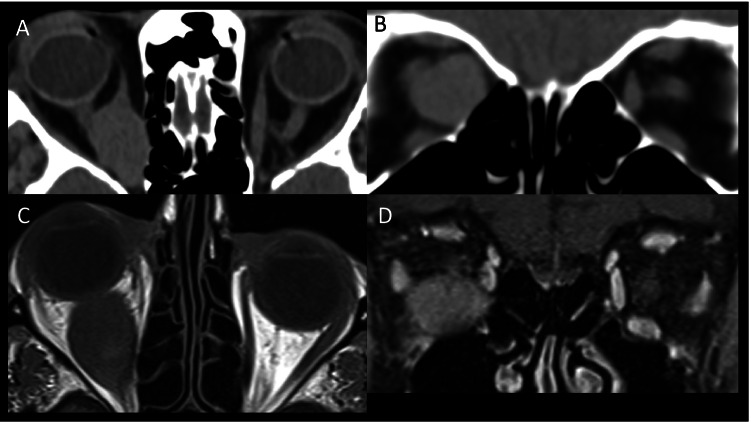
Fig. 9.
Intramuscular lymphoma. Case 1—axial (A) and coronal (B) CT scan showing significant enlargement of the right medial rectus muscle without involving the anterior tendon and causing optic nerve displacement. Case 2—axial T1-weighted MRI (C) shows isointense enlargement of the right inferior rectus muscle and right proptosis. Coronal fat-suppressed contrast-enhanced T1-weighted MRI (D) demonstrates heterogenous enlargement of the right inferior rectus muscle

