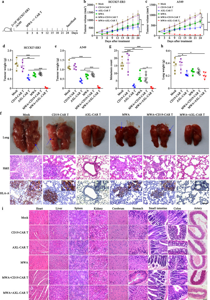
Fig. 3. MWA promotes antitumour activity of AXL-CAR T cells against lung cancer.
a Schematic representation of the experiment examining effects of a combination of MWA and CAR T cell administration (n = 5 mice per group). b/c Tumour volume of mice subcutaneously injected with HCC827-ER3 or A549 cells. d, e Mean tumour weight in each group at the end point. f Detection of HLA-A expression in harvested lung tissues from each group. Representative images are shown (n = 5 per group). Positive staining for HLA-A was observed in lung tissues from mock, CD19 and/or MWA groups. Blue arrow indicated metastatic nodules. g Counted metastasis in lungs of each group. h The weight of lungs from each group. i Histopathological analysis of murine organ tissues. Different tissues were harvested, formalin-fixed, paraffin-embedded, and stained using haematoxylin and eosin (HE) staining. Representative staining image fields (magnification ×400) are shown (n = 5 per group). Scale bars represent 100 μm (f) or 50 μm (i). Data are presented as mean ± SD (b‒e, g, h) and analysed by two-way ANOVA (b, c) or one-way ANOVA (d, e, g, h) with Tukey’s multiple comparisons test. *p < 0.05, **p < 0.01, ***p < 0.001. Source data and exact p values are provided as a Source Data file.

