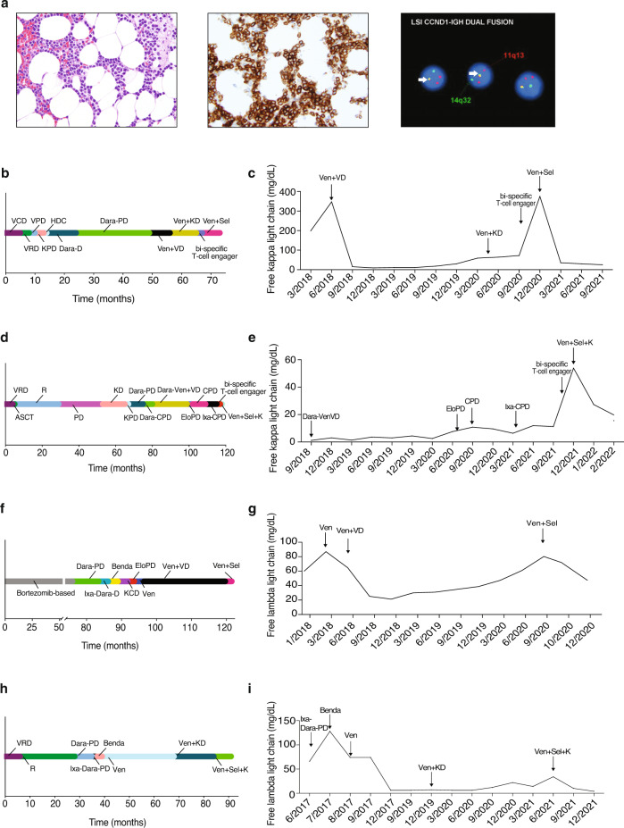
Fig. 1. Clinical response to selinexor and venetoclax in heavily pre-treated t(11;14) multiple myeloma.
Histopathologic findings in bone marrow biopsy at diagnosis showed extensive involvement by plasma cell myeloma. Neoplastic plasma cells were small and mature in appearance and present in a diffuse interstitial distribution comprising ~50–60% of overall marrow cellularity. Hematoxylin and eosin stain, ×400 magnification (a, left). Immunohistochemistry (IHC) for CD138 highlights neoplastic plasma cells present in abnormal clusters, ×400 magnification (a, middle). Fluorescence in situ hybridization (FISH) studies showed CCND1-IGH fusion in 95% of cells (yellow fusion signals indicated by arrow; a, right). Timeline of prior treatments and free light-chain response for Patient 1 (b, c), Patient 2 (d, e), Patient 3 (f, g) and Patient 4 (h, i). V bortezomib, C cyclophosphamide, D dexamethasone, R lenalidomide, P pomalidomide, K carfilzomib, HDC high dose cyclophosphamide, Dara daratumumab, Ven venetoclax, Sel selinexor, Ixa ixazomib, Elo elotuzumab, ACST autologous stem cell transplant, Benda bendamustine.

