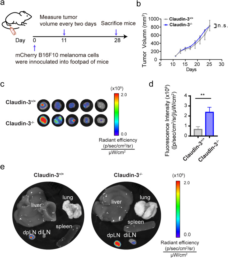
Figure 1.
Loss of claudin-3 augmented B16F10 melanoma lymphatic metastasis. (a) Schematic flowchart shows the establishment of B16F10 melanoma transplantation model in mice. (b) The tumor volume was measured and calculated after day 9 as described in the method part. (ns: no significance) (n = 5 mice per group) (c) mCherry fluorescence was detected by IVIS in ipsilateral popliteal lymph nodes (dpLN) of the footpad injection site from claudin-3−/− and claudin-3+/+ mice. (n = 5 mice per group) (d) Quantification of metastasized mCherry labeled B16F10 cells in dpLN was measured by IVIS. Statistical analysis of fluorescence was conducted. Values are shown as the mean ± SEM. (**p < 0.01) (n = 5 mice per group) (e) After sacrificing the mice, ipsilateral popliteal lymph node (dpLN), ipsilateral inguinal lymph node (diLN), liver, lung, and spleen were collected. Representative images show the fluorescence examined by IVIS.

