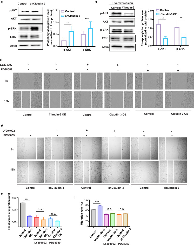
Figure 5.
Knockdown of claudin-3 in SVEC4-10 cell activated the PI3K signaling pathway. (a, b) Western blot analysis was conducted to detect the phosphorylation of AKT and ERK in transfected SVEC4-10 cells as indicated. Total AKT and ERK were detected for normalization. (**p < 0.01, ***p < 0.001) (n = 3 per group) (c) SVEC4-10 Control and OE cells were pretreated with the inhibitor of PI3K (LY294002) (10 μM) or ERK (PD98059) (10 μM). Scratching assay was conducted and the photos were taken under a microscope at the indicated time. Scale bar, 100 µm. (d) SVEC4-10 Control and knockdown cells (shClaudin-3) were pretreated with the inhibitor of PI3K (LY294002) (10 μM) or ERK (PD98059) (10 μM). Scratching assay was conducted and the photos were taken under a microscope at the indicated time. Scale bar, 500 µm. (e) Wound width of result in (c) was quantified and analyzed. (n.s.: no significance; ***p < 0.01) (n = 5 per group) (f) Wound width of result in (d) was quantified and analyzed. (n.s.: no significance; ***p < 0.01) (n = 5 per group).

