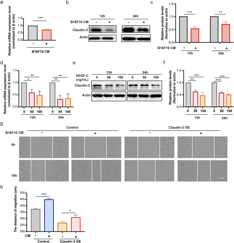
Figure 6.
VEGF-C is sufficient to downregulate claudin-3 expression on SVEC4-10 cells. (a) SVEC4-10 cells were co-cultured with conditional medium (CM) from B16F10 cells for the indicated time. RT-PCR analysis was used to examine the mRNA expression of claudin-3. (***p < 0.001) (n = 3 per group) (b) Western blot analysis indicated the protein expression level of claudin-3 protein in SVEC4-10 cell. (c) Quantitative analysis for western blot results. (**p < 0.01, ***p < 0.001) (n = 3 per group) (d) SVEC4-10 cells were treated with 0 ng/mL, 50 ng/mL or 100 ng/mL VEGF-C for 12 h or 24 h. RT-PCR analysis was used to examine the mRNA expression of claudin-3. (*p < 0.05, **p < 0.01) (n = 3 per group) (e) Western blot analysis indicated the protein expression level of claudin-3 protein in SVEC4-10 cell. (f) Quantitative analysis for western blot results. (**p < 0.01, ***p < 0.001) (n = 3 per group) (g) Wound healing assay of SVEC4-10 cultured with CM from the B16F10 cells and normal control. The ratio of CM to fresh complete medium was 1:3. Photos were taken at the beginning and 16 h later. Scale bar, 100 µm. (h) Wound width was quantified and analyzed by t-test. (*p < 0.05, ***p < 0.001) (n = 5 per group).

