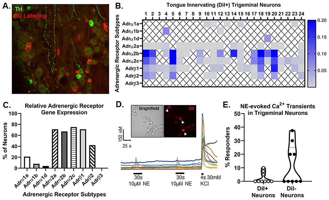
Figure 2. Anti-tyrosine hydroxylase (TH) staining and adrenergic signaling in tongue trigeminal ganglia neurons (TGN).

A) Representative images of retrograde labeling (red) and anti-TH immunoreactivity (green) in the mandibular branch of the TG from an adult female C57Bl/6 mouse 10 days after DiI injection into the tongue. B) Adrenergic receptor subtype gene expression from retrogradely labeled (DiI+) tongue-innervating TG neurons from 2 female C57Bl/6 mice that were manually picked to perform single-cell PCR. Gene expression was calculated for relative mRNA expression for each cell for each gene. Gapdh was used as an internal control. Data are presented as heatmap for each gene for each cell. An X indicated that expression was below the level of detection (CT<38) for that cell. C) Number of DiI+ neurons expressing each gene were counted and presented as percentage of neurons expressing the target gene. D) Representative Ca2+ imaging traces demonstrate the evoked Ca2+ transient in response to 10μM NE applied for 30 sections to a (Inset) mixed field of dissociated TGN containing both retrograde labeled (DiI+, arrow heads) and non-labeled (DiI−) cells. KCl-evoked depolarization (30mM, 4s) was used to establish cell viability in the field. E) Quantitative analysis of the percent of NE-responsive DiI− and DiI+ neurons across 9 mice (5 C57Bl/6, 4 nude). A neuron was considered responsive if NE evoked a Ca2+ transient ≥20% of baseline Ca2+ concentration. At least 15 neurons were assessed per mouse and the percent of responsive neurons per mouse are depicted to representent biological variance.
