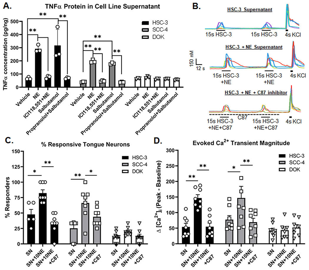
Figure 5. Norepinephrine (NE) stimulation increases cancer cell TNFα secretion and trigeminal ganglia neuron (TGN) activation.

A) Supernatant (SN) from oral cancer (HSC-3, SCC-4) and dysplasia (DOK) were collected 48 h after stimulation with vehicle (0.05% HCl, 0.05% EtOH, 0.02% water), 10μM NE, 1μM ICI118,551 and 10μM NE, 1μM Salbutamol, and 10μM propranolol and 1μM Salbutamol. Blockers were added 1 hour prior to the addition of agonists. Data represent the mean (±SEM) of three different cell passage determinations of TNFα concentration. **p<0.01. B) Example traces of HSC-3 supernatant-evoked (top), 10μM NE-stimulated HSC3 supernatant-evoked (middle) and 10μM NE-stimulated HSC-3 SN in the presence of TNF inhibitor, C87, evoked (bottom) Ca2+ transients. KCl-evoked depolarization (30mM, 4s) was used to establish cell viability. Quantitative analysis was performed to generate (C) percent responders and (D) magnitude of the evoked Ca2+ transient in response to stimulation from SN (n=8 mice), 10μM NE-stimulated supernatant-evoked (SN+NE, n=8 mice) and 10μM NE-stimulated SN in the presence of C87 (SN+10NE+C87, n=8 mice). Transient magnitude was calculated as peak minus baseline Ca2+ concentration. There was no C87 vehicle control (0.02% DMSO) included in the supernatant samples. *p<0.05, **p<0.01
