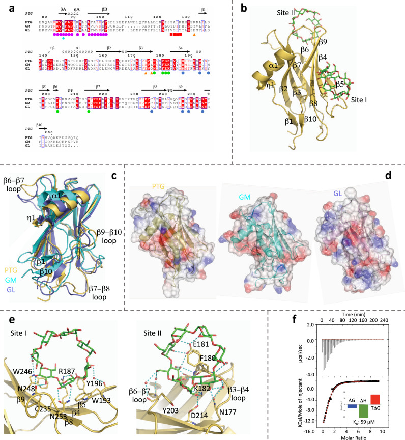
Fig. 1. PTG binding mode to carbohydrates.
a Structural alignment for PTG, GM, and GL. PTG residues interacting with β-cyclodextrin are indicated with blue circles for site I and green circles for site II. PTG ubiquitinated lysines are highlighted with orange triangles, while phosphorylated serine in GM and GL with a cyan diamond. Magenta circles are shown below PP1-interacting residues from the PTG N-terminal region (RVXL-containing signature), while red squares point to the SALK signature. b Crystallographic structure of the PTG CBM21 (yellow) in complex with β-cyclodextrin (green). c Superposition of CBM21 structures of PTG (yellow), GM (cyan), and GL (purple). d Electrostatic surface representation for the three CBM21. e Detailed interaction with β-cyclodextrin at the two binding sites. f ITC returns an enthalpically-driven binding of β-cyclodextrin to the CBM21 with a 1:1 stoichiometry.

