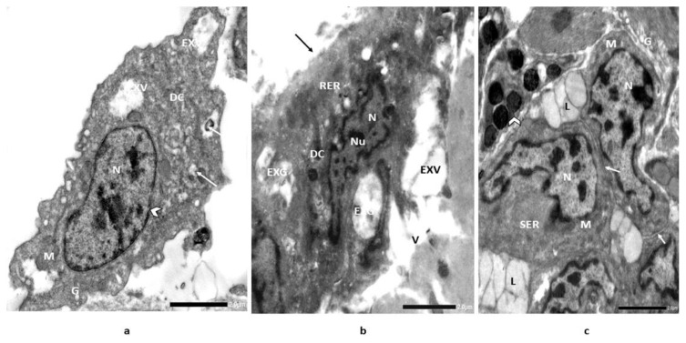
Figure 6.
Electron micrographs illustrate [A] section of digestive glands (control) of snail, Monacha cartusiana (Mǜller) showing nucleus (N), regular nuclear membrane (head arrow), mitochondria (M), execratory vacuoles (EXV), execratory gland (EXG) (arrow), digestive cells (DC) and Golgi bodies (G); [B] section in digestive gland of ZnONPs-treated snail, H. aspersa for 14 d showing destructed cell membrane (arrow), rough endoplasmic reticulum (RER), destructed and irregular execratory glands (EXG), execratory vacuoles (EXV), eccentric nuclei (Nu) with irregular and dilated nuclear membrane (head arrow), nucleus (N) and vacuoles (V) and [C] section in digestive gland of recovered individuals for 14 d showing significant forming of digestive cells (DC) (head arrow), regular cellular membranes (arrow), diluted intercellular spaces, mitochondria (M), smooth endoplasmic reticulum (SER), Nemours shapes of nucleus (N) with irregular nuclear membrane, lipid droplets (L) and slight Golgi bodies (G) [Glutaraldehyde-OsO4 fixed uranyl acetate lead citrate stained preparation 4000X].

