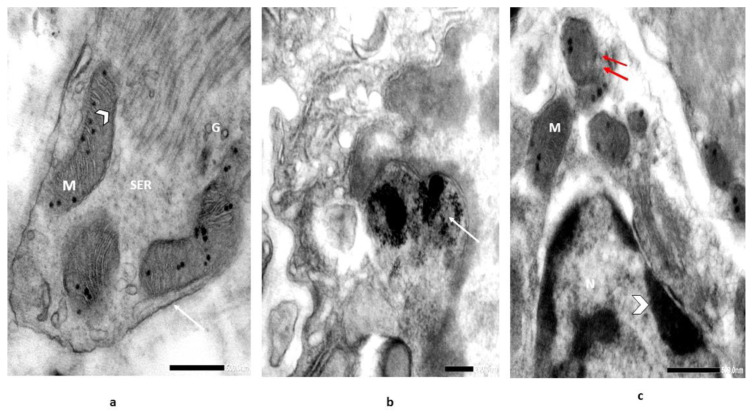
Figure 7.
Electron micrographs illustrate [A] section of digestive g. of snail, Monacha cartusiana (Mǜller) (control) showing normal mitochondria (M) with lamellar cristae (head arrow), smooth endoplasmic reticulum (SER), cell wall (arrow), and Golgi bodies (G); [B] magnified field to obtain NPs precipitation in the lysosome (arrow), lack of mitochondria (M) and rough endoplasmic reticulum (RER), and [C] section in the digestive gland of recovered individuals showing forming of mitochondria (M) with concentric cristae and disrupted mitochondrial membranes (double arrow) and nucleus (N) with migration & aggregation of chromatin is likely to be detectable in the nucleus (head arrow) [Glutaraldehyde-OsO4 fixed uranyl acetate lead citrate stained preparation 10000X].

