FIGURE 5:
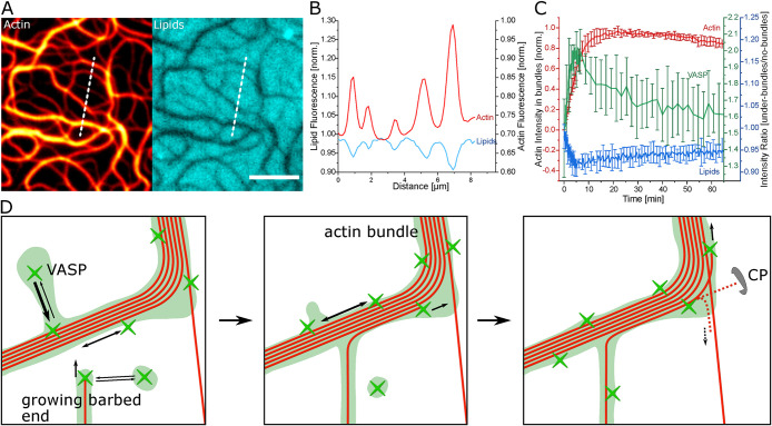
Reorganization of lipid bilayer through VASP accumulation. (A) Lipid fluorescence (Texas Red-DHPE) inversely correlates with actin bundles formed with 0.125 µM VASP. (B) Intensity profile of the bundle cross-section indicated in by the dashed line in (A). (C) Lipid intensity (Texas Red-DHPE) changes reciprocally to the actin intensity during polymerization at 0.25 µM VASP. Afterward it normalizes more toward the values at the start of polymerization inversely to the way the VASP-Alexa532 intensity behaves under bundles. (D) Schematic of the bundle formation mechanism. On the fluid bilayer, patches of VASP can freely diffuse to actin bundles and alongside the bundles. F-actin bundles have more sites to bind VASP and therefore a higher dynamic VASP concentration than single filaments. Barbed ends growing toward bundles are captured and aligned by localized VASP. Filaments are most likely to be continuously polymerized by the higher VASP concentration on the larger bundles, whereas actin filaments polymerizing away from VASP are inhibited by CP. This leads to an enhanced thickening of larger bundles. All scale bars are 5 µm.

