FIGURE 1:
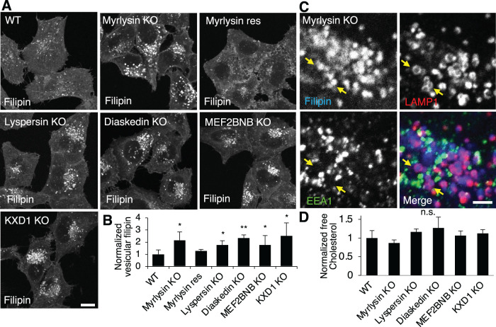
BORC-subunit KO increases lysosomal cholesterol. (A, B) WT, BORC subunit-KO, and myrlysin-rescued (res) HeLa cells were fixed, stained with filipin, and analyzed by confocal microscopy. The intensity of vesicular filipin was quantified (n = 106, 83, 76, 46, 39, 86, and 122) and normalized to the intensity in WT cells. (C) Myrlysin-KO cells were treated as in A and costained with antibodies to EEA1 and LAMP1 for early endosomes and lysosomes, respectively. Part of a cell is shown at high magnification, and LAMP1-wrapped filipin-positive vesicles are pointed by arrows. (D) Cell lysates were extracted from the indicated cells and subjected to enzymatic measurement of free cholesterol. Bar graphs are presented as mean ± SD; p values were determined by Student’s t test. *p < 0.05, **p < 0.01, n.s., not significant (vs. WT). Scale bars, 5 μm.

