FIGURE 1:
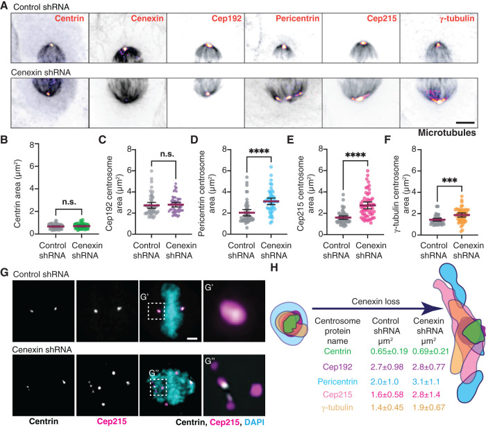
Cenexin is needed for PCM cohesion. (A) Metaphase HeLa cells mitotic centrosomes labeled for centrosome markers: centrin, cenexin, Cep192, pericentrin, Cep215, γ-tubulin (Fire LUT) and MT marker, and α-tubulin (gray). Control shRNA (top) and cenexin shRNA (bottom) -treated cells shown. Scale bar, 5 μm. (B–F) Representative scatter plots depicting two-dimensional areas (μm2) of centrin (B), Cep192 (C), pericentrin (D), Cep215 (E), and γ-tubulin (F) in control and cenexin shRNA-treated metaphase cells. Mean with 95% confidence intervals shown. Unpaired, two-tailed Student’s t tests; n.s., not significant; ***, p < 0.001; ****, p < 0.0001. (G) Control shRNA (top) and cenexin shRNA (bottom) metaphase cell projection. Centrin (gray), Cep215 (magenta), and DNA (DAPI; cyan) shown. Insets magnified 3× from G′ and G″. Scale bar, 5 μm. (H) Model depicting representative centrosome protein outline from a single representative mitotic centrosome reflecting changes resulting from cenexin loss. Mean two-dimensional areas (μm2) ± SD are provided. For graphs: detailed statistical analysis in Supplemental Table S1. See Supplemental Figure S1.

