FIGURE 3:
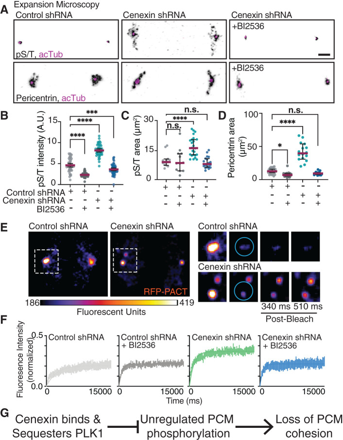
Cenexin is required for PCM cohesion and PLK1 for PCM dispersion. (A) Expansion microscopy of control and cenexin shRNA metaphase cell centrosomes with or without BI2536. Centrosomes immunolabeled for pS/T (inverted gray, top) or pericentrin (inverted gray, bottom), centrioles decorated with acetylated tubulin (acTub; magenta). Scale bar, 5 μm. (B–D) Representative scatter plots depicting pS/T mean intensity at metaphase centrosomes (B), and pS/T (C) and pericentrin (D) expanded two-dimensional areas (μm2) in control shRNA and cenexin shRNA metaphase cells with or without BI2536. Mean (magenta) with 95% confidence intervals displayed. One-way ANOVA with multiple comparisons to control cells; n.s., not significant; *, p < 0.05; **, p < 0.01; ***, p < 0.001; ****, p < 0.0001. (E) FRAP examples of control and cenexin shRNA metaphase cell centrosomes marked with PCM marker RFP-PACT. Magnified insets of boxed centrosomes depict examples of prebleach, bleach, and postbleach time points. Cyan circles represent the region in which a 405-nm laser was applied for photobleaching. Scale bar, 5 μm. (F) FRAP curves for metaphase control shRNA (gray), control shRNA plus BI2536 (black), cenexin shRNA (green), and cenexin shRNA plus BI2536 (blue) cells. SEM for n > 6 cells. (G) A proposed model that cenexin binds and sequesters PLK1 inhibiting PLK1’s potential to have unregulated phosphorylation on PCM substrates that results in PCM cohesion loss. For graphs: statistical analysis in Supplemental Table S1. See Supplemental Figure S3.

