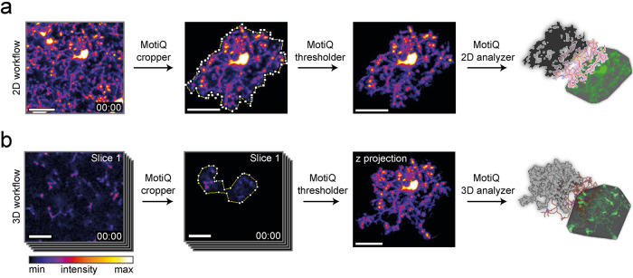
FIGURE 1:
MotiQ workflow. MotiQ offers two main approaches for microglial cell analysis: (a) 2D time-lapse image analysis and (b) analysis of 3D time-lapse image stacks. Both workflows are composed of three steps: single-cell selection with MotiQ cropper, image segmentation with MotiQ thresholder, and cell quantification using (a) MotiQ 2D analyzer or (b) MotiQ 3D analyzer. Cell volume, cell surface, cell skeleton, and the convex hull of the cell are reconstructed and analyzed and serve as a basis for the calculation of more than 60 parameters of microglial morphology, dynamics, and fluorescence kinetics. MotiQ automatically generates 3D image visualizations by implementing Volume Viewer (ImageJ plug-in by Kai Uwe Barthel, Internationale Medieninformatik, HTW Berlin, Berlin, Germany). Scale bars, 20 µm.

