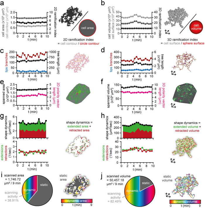
FIGURE 2:
Analysis of in vivo two-photon microscopy data. Selected morphological and dynamic parameters of a representative cortical microglial cell in CX3CR1GFP/wt mice imaged with in vivo two-photon microscopy and analyzed with MotiQ: (a) 2D ramification index, (b) 3D ramification index, (c, d) number of tips, number of branches, and process tree length, (e, f) spanned area or spanned volume and cell polarity vector length (5× magnified for better visualization), (g, h) shape dynamics and number of extensions and retractions as parameter for cell shape alteration, and (i, j) scanned area or scanned volume represent the brain area or brain volume that has been occupied by a microglial cell over a selected time span (here 9 min).

