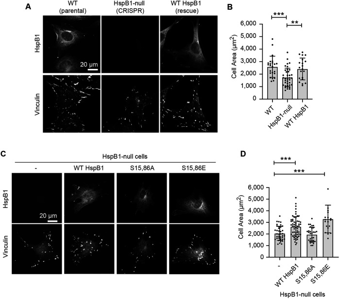
FIGURE 4:
HspB1 affects cell spreading in a phosphodependent manner. (A) immunofluorescence microscopy of cells spread on glass coverslips coated with 10 µg/ml fibronectin. Subcellular distribution of HspB1 (top row, cytoplasmic) and vinculin (bottom row, FA) in WT and HspB1-null cells, and in null cells expressing the WT HspB1 rescue construct. (B) Graph of cell area measurements shows the decreased cell spread in HspB1-null cells is rescued by expressing WT HspB1 rescue construct. (C) immunofluorescence localization of HspB1 (top row) in HspB1-null cells, and in null cells expressing the rescue constructs for WT HspB1 and nonphosphorylatable S15,86A HspB1 and phosphomimetic S15,86E HspB1. Vinculin immunofluorescent localizations in same cells (bottom row). (D) Graph of cell area measurements show increased cell spreading in cells expressing WT and S15,86E HspB1, but no difference between HspB1-null cells and cells expressing S15,86A HspB1. Scale bar of 20 microns for widefield fluorescent images. Graphs are mean with standard deviations and unpaired t tests were used to determine p-values of **p < 0.01, ***p < 0.001.

