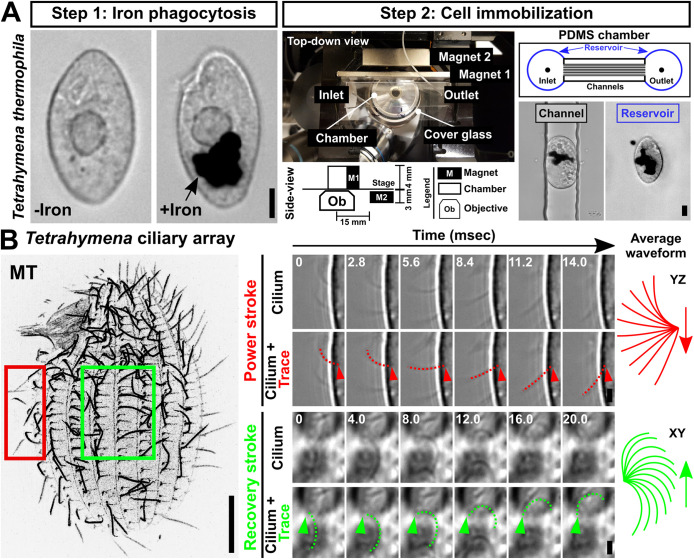
FIGURE 2:
Tetrahymena live cell immobilization technique. (A) DIPULL microscopy setup. Step 1: Tetrahymena cells are fed iron particles. Cell images pre- and post–iron engulfment. Step 2: Cells are introduced into a microfluidic chamber and immobilized via a constant external magnetic field. To track intracellular dynamics, imaging was performed on cells that were trapped within channels (black outline). The visualization of extracellular dynamics was performed on cells that are trapped within the chamber reservoir (blue outline). Scale bars, 10 µm. (B) Visualization of ciliary dynamics via DIPULL-immobilized live Tetrahymena cells. Left panel: Tetrahymena ciliary array. Bar, 10 µm. Right panel: Time-lapse images of power and recovery strokes. Time intervals (ms) are indicated. Dotted lines mark manual cilia traces. Average power stroke, red; average recovery stroke, green. Scale bars, 2 µm.

