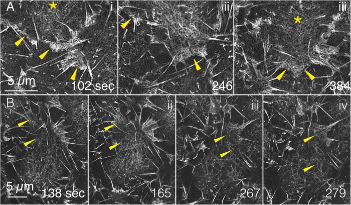
FIGURE 2:
GI-SIM shows that medioapical arrays are continuous with lamellipodia. F-actin labeled with GFP-MoeABD. (A) Three panels are magnified ROIs from Supplemental Movie 1 that show medioapical arrays (yellow asterisks) where they merge with the lamellipodia (yellow arrowheads). (B) Four additional panels (from a different embryo) also demonstrate continuity between medioapical arrays and lamellipodia. The top panel in Supplemental Movie 5 includes each of the time points shown in the top row of Figure 2; the bottom panel in Supplemental Movie 5 shows time points 120–330 s.

