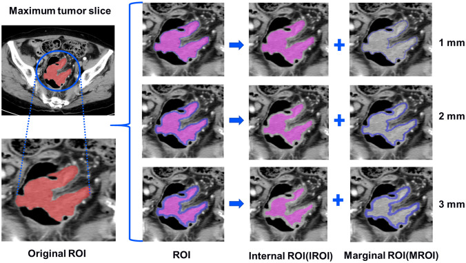
Figure 3.
The tumors were segmented on venous phase, three groups of different marginal and internal regions were formed by retracting the tumor margin by 1, 2, and 3 mm on the representative section. The red areas represent the maximum slice of the tumor (ROI). The blue areas represent the rings with the thickness of 1-3mm in the marginal regions of the tumor (MROI), respectively. The hot-pink areas represent the remaining internal regions of the tumor (IROI) after removing the rings, respectively. ROI, regions of interest. MROI, the marginal areas of tumor. IROI, the interior areas of tumor.

