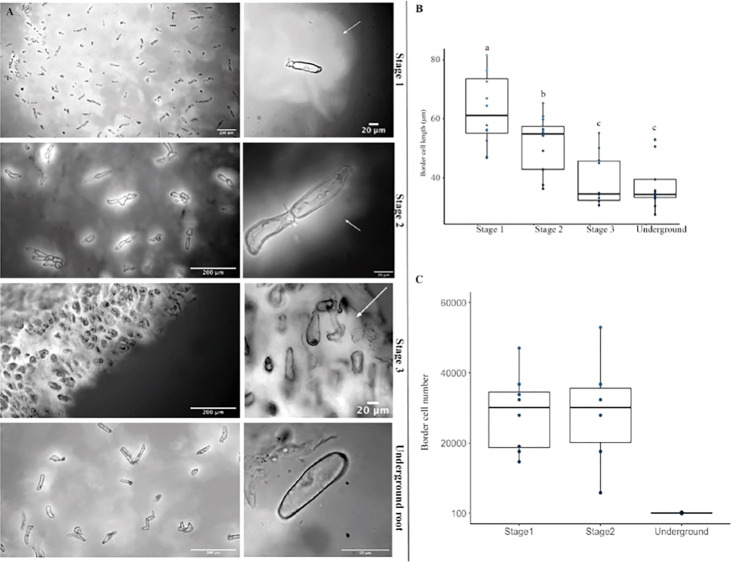
Figure 6.
The mucilage produced in the aerial root surface is enriched in border cells, and the border cell secretes mucilage. (A) The left panel shows a general view of border cells embedded in its mucilage in stages 1, 2, and 3, and the underground root system. The right panels are magnified images of a light microscope representing the shape of the Ames 19897 accession border cells (GRIN) from aerial roots and underground roots. (B) Border cell lengths from aerial root stages 1 and 2 are larger than stage 3 and underground border cells. (C) Stages 1 and 2 have more border cells than stage 3. Error bars represent the standard deviation between biological replicates counted in two independent experiments. Analysis of variance (ANOVA) was performed on border cell length, and number values, and significance difference represents “a” p < 0.05; “b” p<0.01, “c” p<0,001.

