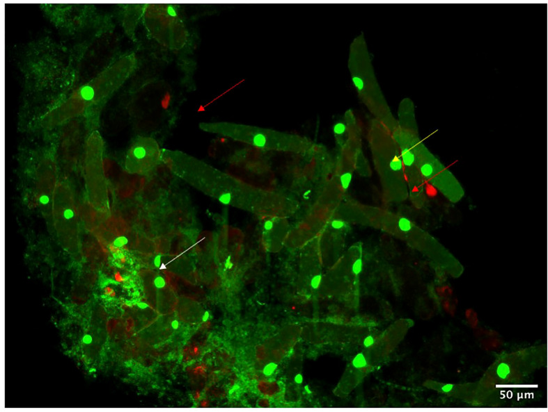
Figure 7.
Root Border Cells from maize detach from the aerial root cap and remain in the mucilage environment. Fluorescent microscopy of border cells stained with propidium iodide marks dead cells (red arrows), and acridine orange marks viable cells (yellow arrows). White arrows indicate bacterial colonies trapped or attached to the border cells.

