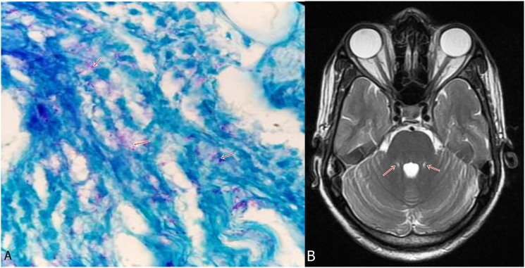
Figure 2.
A 40-year-old man presented with sensory motor polyneuropathy, lower motor neuron facial palsy and thickened bilateral peroneal nerves. He also had an erythematous hypo-anesthetic patch on his face. There was no suggestion of appendicular or gait ataxia. Sural nerve biopsy demonstrated lepra bacilli (A, arrows) (Wade Fite stain, x1000). MRI brain demonstrated bilateral symmetrical hyperintensities in middle cerebellar peduncles (B, arrows). (Case 1).

