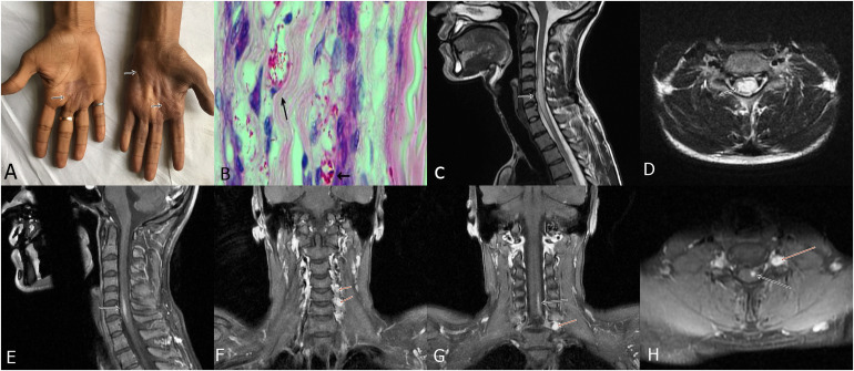
Figure 3.
A 28-year-old man presented with multiple anesthetic patches on the body, weakness of both the upper limbs and clawing. Ulnar nerves were thickened. Hyperpigmented and anesthetic skin lesions were present on the hands along with wasting of hand muscles (A, arrows). Sural nerve biopsy demonstrated lepra bacilli forming globi (Wade Fite stain, x1000) (B, arrows). MRI of the spine demonstrated a long segment hyper intensity along with cord swelling (myelitis) in the cervical region (C, arrow). Axial cuts showed involvement of the central spinal cord (D, arrow); post-contrast image showed enhancement of the lesion (E, arrow). Coronal post-contrast images depicted enhancement of the dorsal root ganglion (Ganglionitis) (F, arrows). Coronal and axial post contrast image demonstrated contrast enhancement of spinal cord lesion as well as ganglion (G and H, arrows). (Case-2).

