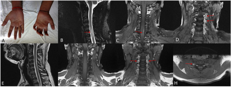
Figure 4.
A 28-year-old man presented with polyneuropathy, thickened nerves and trophic ulcers. Claw hand deformity was present (A). MRI cervical spine T2 sagittal (B) and post contrast coronal (C and D) showed hyperintensity in the cervical spinal cord with contrast enhancement (myelitis) along with contrast enhancement of dorsal root ganglion (ganglionitis). MRI was repeated after 6 months, which showed resolution of myelitis (E and F) but persistence of ganglionitis (G and H). (Case 3).

