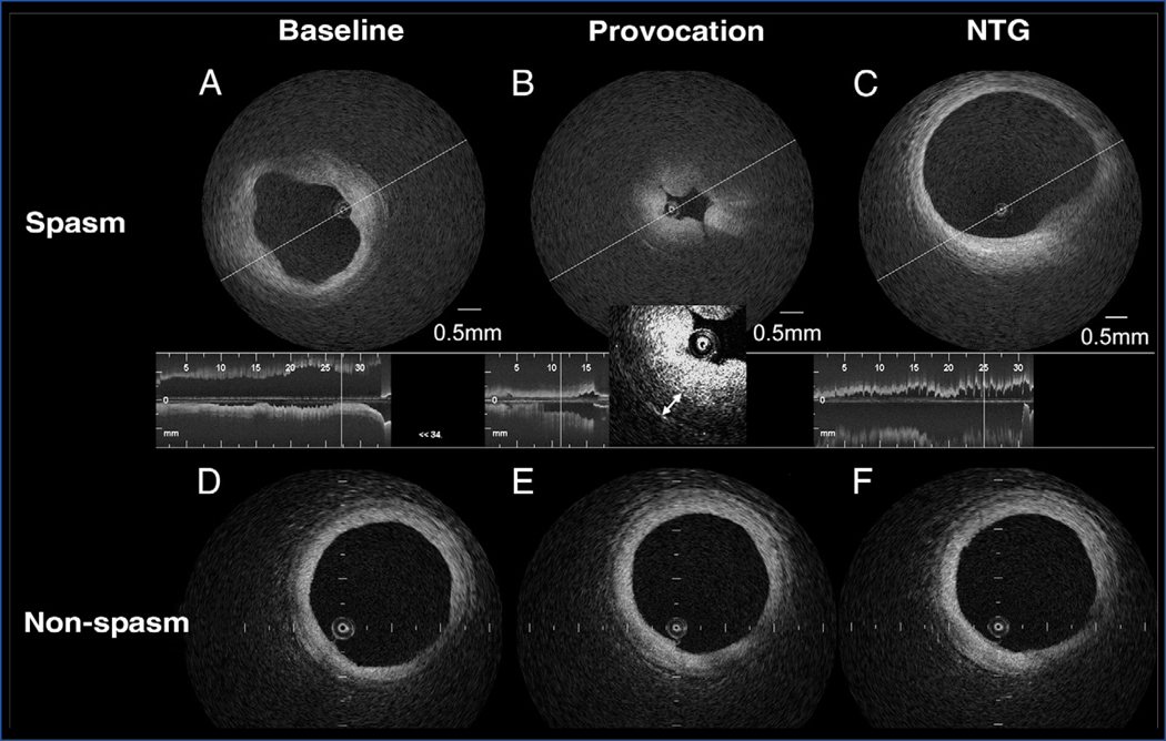
Figure 3. Optical coherence tomography features of vasospastic angina.
Representative intracoronary optical coherence tomography images of a patient with vasospastic angina at a site of coronary vasospasm (top row) versus a site without coronary vasospasm (bottom row). (A) Both intimal bumps around the lumen and a thickened medial layer can be seen at baseline. (B) With provocation, intimal gathering appears, with a further increase in the medial layer (double headed arrows). (C) Both intimal bumps and gathering disappear with intracoronary nitroglycerin (NTG). Compared to the morphology at (D) baseline, no changes are seen at the non-spasm site with either (E) acetylcholine or (F) nitroglycerin administration. Reproduced from reference [75], with permission from Elsevier.

