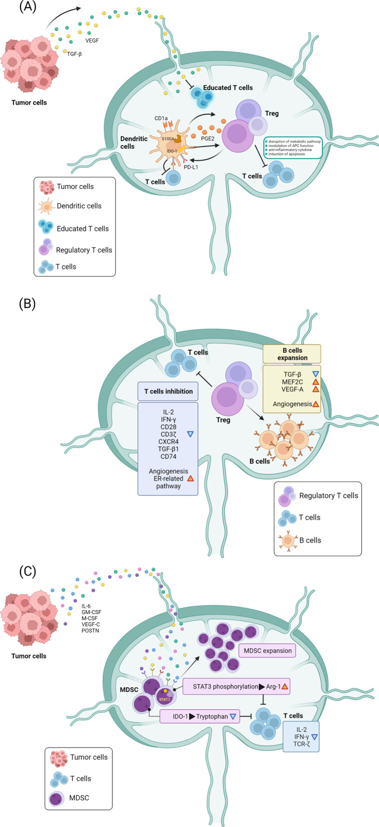
Fig. 1.

Immune microenvironment modulation in the SLN during cancer progression. A Tumor cell-derived soluble factors including TGF-β and VEGF impair T cell and lymphocyte activation by DCs. Expression of COX2 and IDO-1 in DCs also contribute to the expansion of Treg cells which directly diminish T cell activity. Treg cells stimulate a feedback loop to enhance PD-L1 expression on DCs, leading to immune suppression of T cells via the receptor PD-1. S100 and CD1a elevated in the DCs in the SLN also modulate antigen presentation of DCs. B After expansion, Treg cells enter the SLN via HEV and lymphatic vessels. Treg cells inhibit T cell activity, decrease pivotal molecules such as IL-2, IFN-γ, CD28, CD3-ζ chain, CXCR4, TGF-β1 and CD74 to suppress the proliferation, activation and differentiation of T cells. Treg cells also induce B cells expansion and enhance MEF2C expression to alter the proliferation and survival of B cells. In addition, Treg cells promote the estrogen receptor-related pathways in T cells to facilitate angiogenesis and lymphangiogenesis. (C) MDSCs are recruited to the SLN and are activated by external signals such as IL-6, GM-CSF, M-CSF, VEGF-C and POSTN released from distant tumor cells. STAT3 phosphorylation in MDSCs stimulate the production of Arg-1 to compete the essential amino acid with T cells and to suppress T cell function. In addition, IDO-1-expressing MDSCs catalyze the metabolism of tryptophan to kynurenine, a metabolite which induces T cell apoptosis
