Fig. 3.
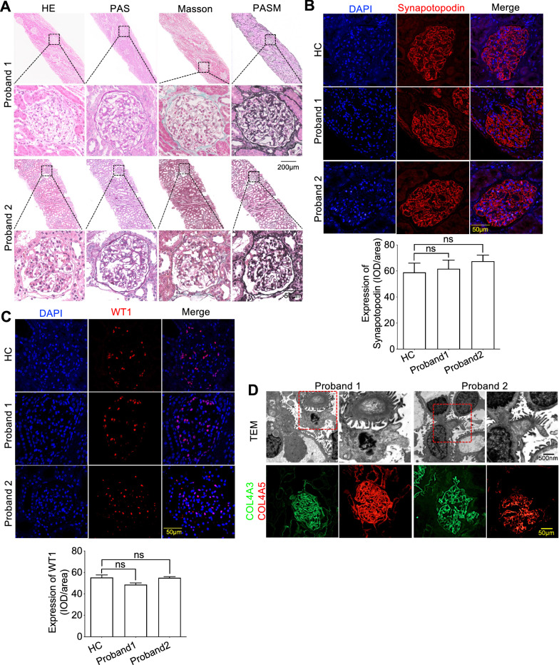
Compound heterozygous variants locating after vitB12-binding domain of CUBN exhibited normal GFB. A hematoxylin–eosin (HE), periodic acid–Schiff (PAS), periodic acid-silver metheramine (PASM) and Masson of kidney biopsy of the two probands showed there was no obvious proliferation of glomerular mesangial cells, inflammatory cell infiltration, fibrosis, glomerular sclerosis or segmental sclerosis. B and C Immunofluorescence staining showed the expression of podocyte membrane marker Synapotopodin and nuclear marker WT1 was normal as compared to HC. D Representative photomicrographs by transmission electron microscopy (TEM) analyses showed there were no thickening of GBM or widening and fusion of podocytes and immunofluorescence staining with COL4A3 and COL4A5, the important components of GBM, indicated that there was no exact immune complex deposition. Each group was tested in triplicate, and the data are presented as the mean ± S.D. ns, no significant

