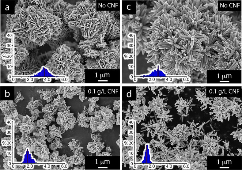
Figure 2.
Micrographs of ZnO particles referred to as flowers a and b (run 1 and run 3 (Table 1)) and sea urchins c and d (run 4 and run 6 (Table 1)) after calcination at 400 °C. The effect of performing the synthesis in the presence of 0.1 g/L of CNF is demonstrated. The histograms show the distribution of particle diameters in micrometers for the flower-shaped particles (a: average size 3.52 μm and b: average size 1.76 μm) and the sea urchin particles (c: average size 2.45 μm and d: average size 1.34 μm) consisting of nanosheets and hexagonally faceted rods, respectively. The red lines represent the average particle sizes. All particles (regardless if synthesized with CNF) were calcinated for accurate comparison.

