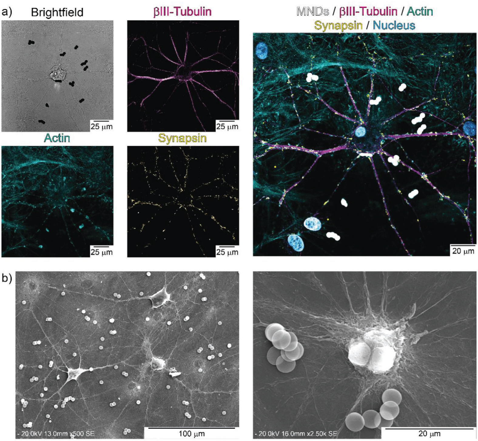
Figure 6.

MMDs colocalization within primary cortical neural networks. a) Confocal images of cortical neurons. Cell cytoskeleton microtubules are stained with anti-Tubulin β-III (magenta), actin fibers are stained with Actin-stain 488 Phalloidin (cyan), presynaptic vesicles are labeled with synaptophysin antibody (yellow), nuclei counterstaining was performed with DAPI (blue), MMDs were imaged in bright field (black), and the color was inverted in the merged image (white). b) SEM image of cortical neural networks exposed to MMDs.
