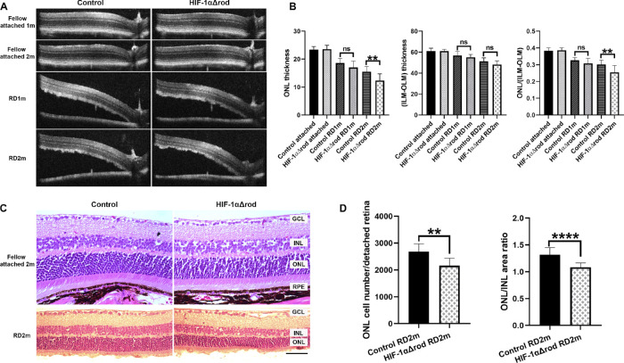
Figure 3.
Conditional knockout of HIF-1α in rods increased photoreceptor cell loss following RD. (A) Optical coherence tomography (OCT) images of fellow attached and detached retinas at 1 and 2 months post-RD in HIF-1αΔrod and control mice. (B) Graphs show the measurements obtained from A. The thickness of ONL and the thickness from the inner limiting membrane (ILM) to the outer limiting membrane (OLM) were measured at 500 pixels away from the optic nerve head using ImageJ. The ONL/(ILM-OLM) ratio was calculated. (C) Hematoxylin and eosin staining of retinal sections of fellow attached and detached retinas at 2 months post RD in HIF-1αΔrod and control mice. Scale bar = 50 µm. (D) Graphs show the measurements obtained from C. The nuclei count in the ONL per detached retina from the optic nerve head to the tip of retinal periphery was measured using ImageJ. The areas of ONL and INL were also measured and ONL/INL area ratio was calculated. Control group N = 9; HIF-1αΔrod group N = 8. Data are shown as mean ± S.D. ns, not significant. **P < 0.01, ****P < 0.0001 (unpaired Student's t-test).

