Figure 2.

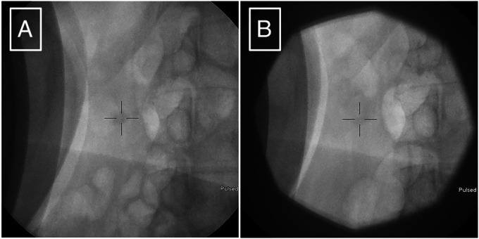
Fluoroscopy obtained during ESWL showing (A) pre-ESWL pancreatolithiasis, and (B) post-ESWL pancreatolithiasis is fractured into smaller fragments. ESWL, extracorporeal shock wave lithotripsy.

Fluoroscopy obtained during ESWL showing (A) pre-ESWL pancreatolithiasis, and (B) post-ESWL pancreatolithiasis is fractured into smaller fragments. ESWL, extracorporeal shock wave lithotripsy.