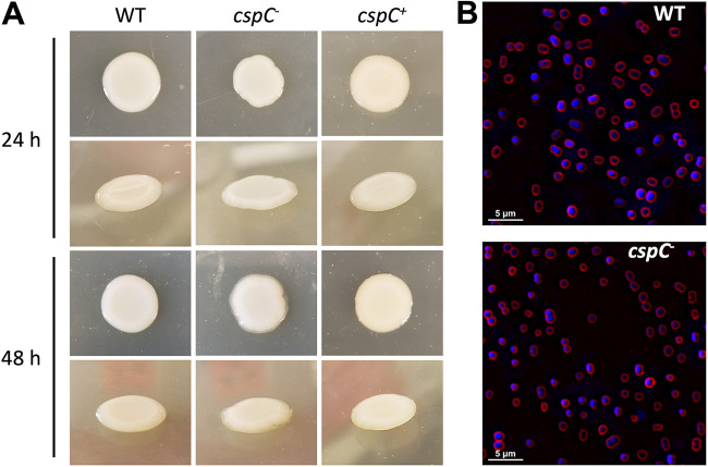
FIG 3.
cspC mutants display a highly mucoid colony phenotype but unaltered cellular morphology. (A) Wild-type (WT), cspC–, and cspC+ strains were grown on LB agar supplemented with hygromycin at 37°C for 24 h (top) and subsequently left to grow for an additional 24 h at room temperature (bottom). Images are representative of 3 experimental repeats. (B) Fluorescence microscopy was used to visualize cell morphology for the wild-type and cspC mutant strain. Cells harvested from bacterial colonies were stained with FM4-64 and DAPI to visualize cell membranes (red) and DNA (blue), respectively. Images are representative of three biological replicates.

