FIG 4.
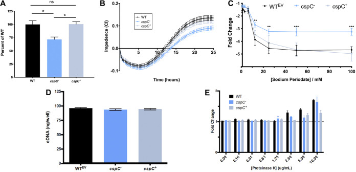
Disruption of cspC results in impaired biofilm formation and enhanced resistance to sodium periodate. (A and B) Comparison of biofilm formation using crystal violet staining following 24 h growth (A) and continuously using an xCELLigence RTCA (B) over a 25-h period. Assays were performed in biological triplicate with 3 technical replicates each. Assays in panel A are corrected for subtle OD600 variations between the wild-type and mutant/complement strains after 24 h of growth under static conditions (Fig. S2). Student’s t test was used to determine statistical significance compared to the wild type. (C) For the biofilm inhibition assay, biofilms were seeded with increasing concentrations of sodium periodate and incubated for 24 h, with the resulting biofilms measured using a crystal violet assay. Data points are from 10 biological replicates and 3 technical replicates. The fold change is reported relative to no-treatment controls for each strain. A two-way analysis of variance (ANOVA) was used to determine statistical significance from the wild type. (D) Extracellular DNA production within the biofilm matrix was measured using a PicoGreen assay kit after 24 h of formation. (E) Biofilms were seeded with increasing concentrations of proteinase K and then measured using a crystal violet assay after 24 h of formation. Change is reported as the fold difference from the no-treatment control. Assays were performed in biological triplicate; Student’s t test determined no statistically significant differences between strains for panels D and E. Error bars are shown as the ±SEM. **, P ≤ 0.01; ***, P ≤ 0.001; ns, not significant.

