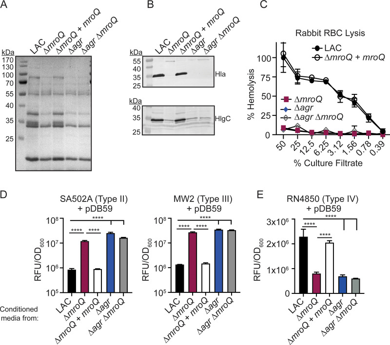
FIG 2.
MroQ contributes to Agr type I and IV activation. (A and B) TCA-precipitated exoproteins (A) and Hla and HlgC immunoblots (B) from LAC, ΔmroQ, ΔmroQ + mroQ, Δagr::tet, and Δagr::tet ΔmroQ strains. (C) Rabbit red blood cell lysis of cell-free culture filtrates derived from LAC, ΔmroQ, ΔmroQ + mroQ, Δagr::tet, and Δagr::tet ΔmroQ strains. (D) pDB59 reporter activity (relative fluorescence units [RFU]/OD600) in SA502A (left) and MW2 (right) upon addition of conditioned medium from LAC, ΔmroQ, ΔmroQ + mroQ, Δagr::tet, and Δagr::tet ΔmroQ strains. (E) pDB59 reporter activity (RFU/OD600) in RN4850 (type IV) upon addition of conditioned medium from LAC, ΔmroQ, ΔmroQ + mroQ, Δagr::tet, and Δagr::tet ΔmroQ strains. Hemolysis and GFP reporter assay data are from one of at least three experiments conducted in triplicate. Immunoblots and GelCode blue-stained gels are a representative of at least four replicates. Means ± SD are shown (n = 3). ****, P < 0.0001 by one-way analysis of variance (ANOVA) with Tukey’s posttest.

