FIG 1.
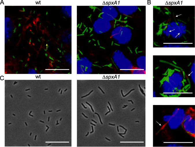
Microscopy of L. monocytogenes ΔspxA1 during intracellular and extracellular growth. (A) L2 murine fibroblasts were infected for 10 h with wt or ΔspxA1 cells and fluorescently labeled to visualize DNA (blue), L. monocytogenes (green), and host actin (red). The yellow arrow indicates a canonical actin comet tail. Images were taken using a 100× objective. (B) Additional examples of cells infected with the ΔspxA1 mutant. White arrows indicate elongated bacteria with disorganized or nonpolar actin recruitment. (C) Phase-contrast images of wt and ΔspxA1 cells grown to early exponential phase in rich anaerobic broth. Bacteria were imaged with a 40× objective. All scale bars represent 10 μm, and all images represent three biological replicates and at least 10 fields of view.

