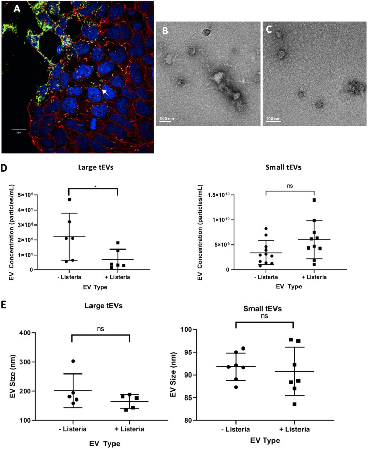
FIG 1.
Extracellular vesicles from L. monocytogenes-infected TSCs. (A) Trophoblast stem cells (TSCs) from C57BL/6 mice were infected with GFP-expressing L. monocytogenes at a multiplicity of infection (MOI) of 100:1. At 24 h postinfection, the cells were fixed and stained with DAPI (blue) and rhodamine phalloidin (Red) which bind to DNA and polymerized actin, respectively. The cells were later imaged with an Olympus FluoView scanning confocal light microscope. The scale bar is 20 μm. (B, C) Transmission electron microscopy images of L-tEVs (B) and S-tEVs (C) from L. monocytogenes-infected TSCs. (D, E) TSC-derived EVs were analyzed by nanoparticle tracking analysis that gives the concentration and size distribution of the nanoparticles. The concentration (D) and mean size (E) of the tEVs with and without infection are given. Comparisons were conducted using Student’s t test; *, P < 0.05; ns, not significant.

