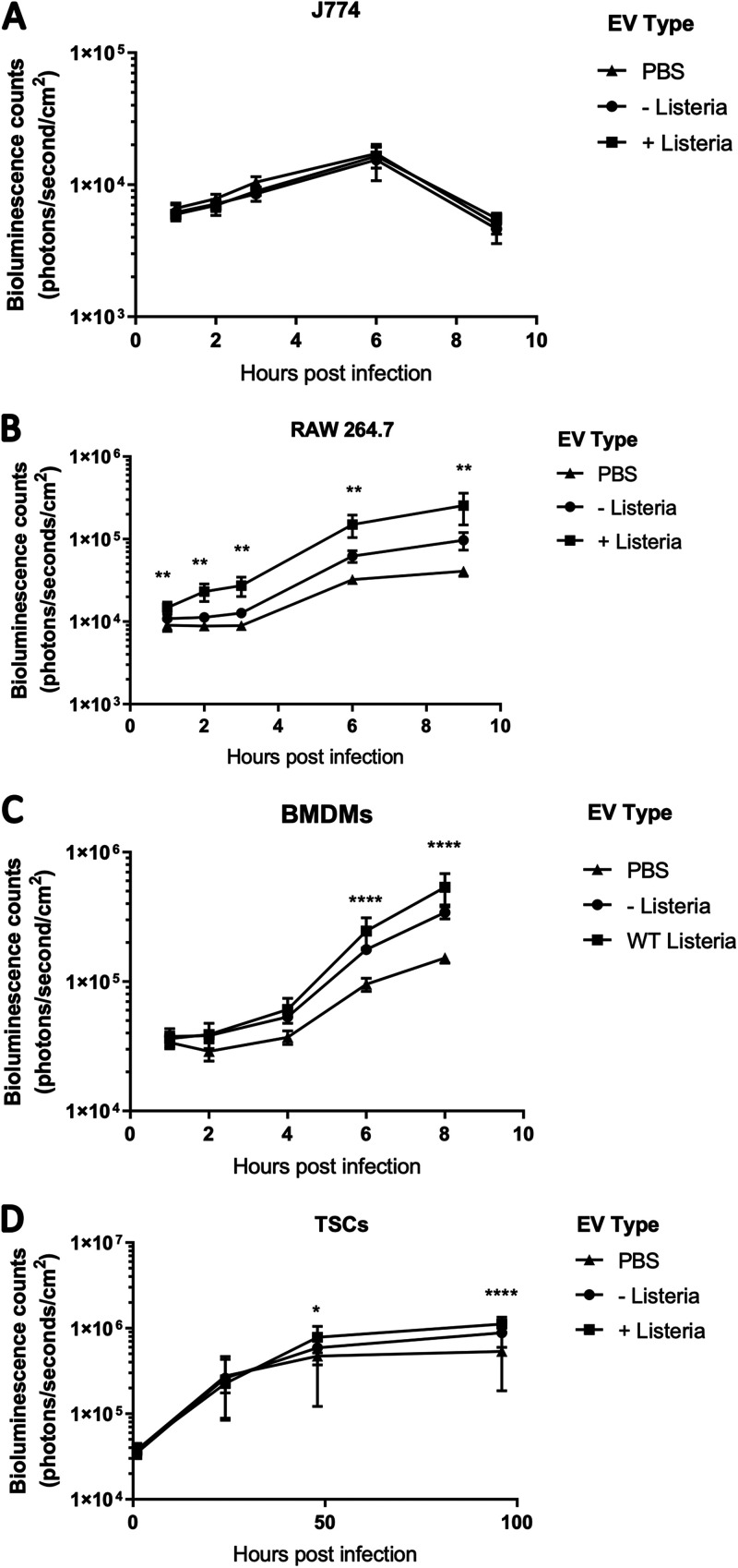
FIG 3.
tEVs from L. monocytogenes-infected TSCs make cells susceptible to L. monocytogenes infection. (A to D) A total of 5 × 10⁵ J774 cells (A), RAW 264.7 cells (B), BMDMs (C), and TSCs (D) were treated with 5 × 10⁶ S-tEVs. After 24 h, the cells were infected at an MOI of 10 (A, B, C) or an MOI of 100 (D) with mid-log phase bioluminescent L. monocytogenes. The cells were imaged using the PerkinElmer in vivo imaging system (IVIS). Each group consisted of six replicates. At each time point, the EV groups were compared using Tukey’s post hoc multiple-comparison test. The stars indicate the any statistical difference between the + Listeria and PBS groups; *, P < 0.05; **, P < 0.01; ****, P < 0.0001.

