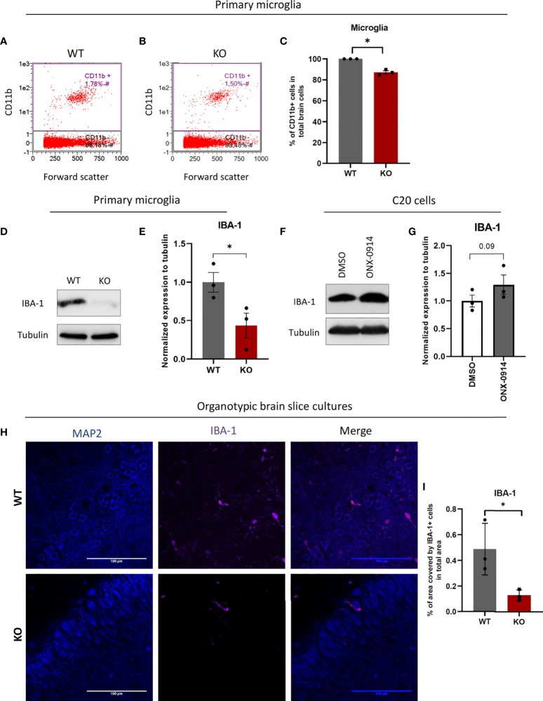
Figure 7.
β5i/LMP7 impairment results in less microglia population in mice brains and differs expression levels of microglial surface markers. After brain dissociation, cell suspensions were stained with CD11b FACS antibody to determine microglia amount in the brain by flow cytometry. (A–C) Flow cytometric analysis of CD11b+ cells in WT (A) and β5i/LMP7 KO (B) mice brains, and comparison analysis of CD11b+ cells (C). (D) Western blot analysis of ionized calcium-binding adapter molecule 1 (IBA-1) in primary microglia. (E) Quantification analysis of IBA-1 Western blot data from (D). (F) Western blot analysis of IBA-1 in C20 cells treated with 200nM of ONX-0914 for 24h. (G) Quantification analysis of IBA-1 Western blot data from (F). (H) Immunofluorescence microscopy analysis of MAP2 and IBA-1 in organotypic brain slice cultures from WT and β5i/LMP7 KO mice brains. (I) Analysis of % area covered by IBA-1+ cells in total area. All data are given (n=3) by mean ± SEM; *: P < 0.05.

