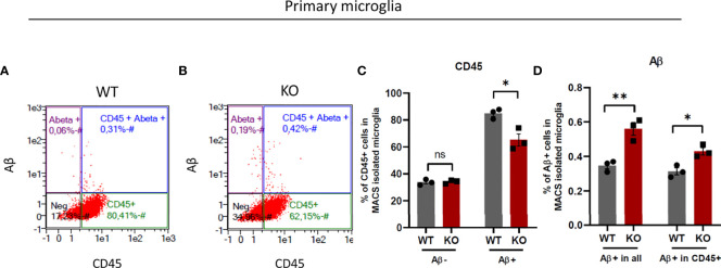
Figure 8.
β5i/LMP7 deficient microglia exhibit more phagocytosis activity. (A–D) Phagocytosis activity was determined by the analysis of TAMRA-Aβ+ cell population in primary micrgolia. (A, B) Gating strategy of flow cytometry analysis of MACS isolated WT (A) and β5i/LMP7 KO (B) microglia incubated with 2µM TAMRA-labeled amyloid-beta (Aβ) toxic oligomers. (C) Bar graphs showing quantification of total CD45+ cells before Aβ treatment, total CD45+ cells after Aβ treatment, (D) Bar graphs showing quantification of total Aβ+ cells and Aβ+ cells in CD45+ cells in primary microglia. All data are given (n=3) by mean ± SEM; *: P < 0.05, **: P < 0.01.

