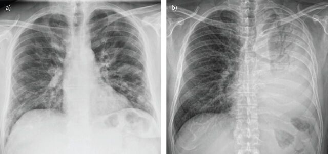
FIGURE 3.
Viral versus bacterial pneumonia. a) Pneumonia due to influenza A virus in a 53-year-old man with cough and dyspnoea. Chest radiography shows bilateral nodular opacities and patchy, focal areas of consolidation, predominantly located in the lower lobes. b) Pneumonia due to Streptococcus pneumoniae in an HIV-infected, 55-year-old man. Chest radiography reveals an extensive consolidation of the left lung with air bronchogram.

