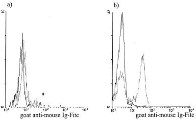
FIG. 4.
Localization of the CD4—anti-CD4–PE MAb complex with goat anti-mouse Ig–FITC. The experiment was performed as outlined in Fig. 3. Goat anti-mouse Ig–FITC was used to stain cultures harvested following overnight culture to assess the presence of the CD4—anti-CD4–PE MAb complex on the cell surface; monocytes and lymphocytes were electronically gated based on their respective forward- versus side-scatter characteristics. The goat anti-mouse Ig–FITC signal was assessed for untagged (——) and CD4-tagged (······) monocytes (a) and lymphocytes (b) (n = 3). The asterisk indicates a goat anti-mouse Ig–FITC signal most likely due to the presence of contaminating CD4+ T cells in the monocyte gate.

