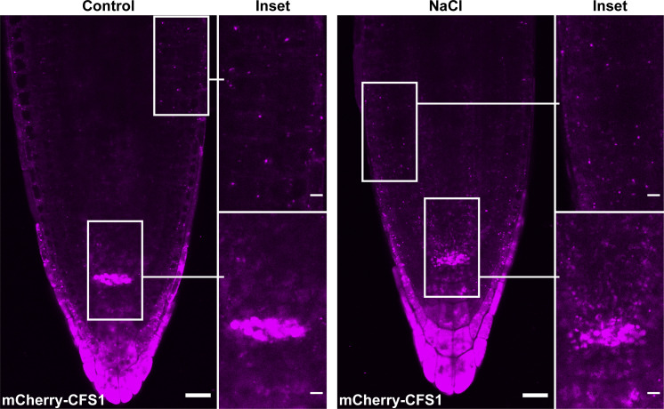
Figure 10.
CFS1 accumulates at the quiescent center cells of A. thaliana. Confocal microscopy images showing that pUBQ::mCherry-CFS1 strongly accumulates at the quiescent center cells of A. thaliana. 5-d-old Arabidopsis seedlings were incubated in either control or 150 mM NaCl-containing 1/2 MS media for 1 h before imaging. Representative images of three replicates are shown. Area highlighted in the white-boxed region was further enlarged and presented in the inset panel. Scale bars, 20 μm. Inset scale bars, 5 μm.

