Figure 5.
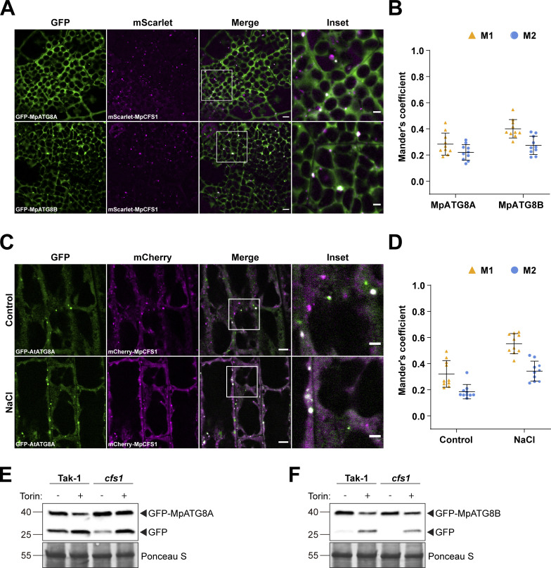
CFS1 function is conserved in M. polymorpha. (A) Confocal microscopy images of M. polymorpha thallus cells co-expressing pEF1::mScarlet-MpCFS1 with either pEF1::GFP-MpATG8A or pEF1::GFP-MpATG8B. 2-d-old thalli were incubated in 1/2 Gamborg B5 media before imaging. Representative images of 10 replicates are shown. Area highlighted in the white-boxed region in the merge panel was further enlarged and presented in the inset panel. Scale bars, 5 μm. Inset scale bars, 2 μm. (B) Quantification of confocal experiments in A showing the Mander’s colocalization coefficients between mScarlet-MpCFS1 and GFP-fused MpATG8A or MpATG8B. M1, fraction of the GFP-fused MpATG8A or MpATG8B signals that overlaps with mScarlet-MpCFS1 signal. M2, fraction of mScarlet-MpCFS1 signal that overlaps with GFP-fused MpATG8A or MpATG8B signals. Bars indicate the mean ± SD of 10 replicates. (C) Confocal microscopy images of Arabidopsis root epidermal cells co-expressing pUBQ::mCherry-MpCFS1 and pUBQ::GFP-AtATG8A. 5-d-old seedlings were incubated in either control or 150 mM NaCl-containing 1/2 MS media for 1 h before imaging. Representative images of 10 replicates are shown. Scale bars, 5 μm. Inset scale bars, 2 μm. (D) Quantification of confocal experiments in (C) showing the Mander’s colocalization coefficients between mCherry-MpCFS1 and GFP-AtATG8A under control or salt-stressed (NaCl) conditions. M1, fraction of the GFP-AtATG8A signal that overlaps with the mCherry-MpCFS1 signal. M2, fraction of the mCherry-MpCFS1 signal that overlaps with the GFP-AtATG8A signal. Bars indicate the mean ± SD of 10 replicates. (E) GFP cleavage assay of pEF1::GFP-MpATG8A in M. polymorpha wild type (Tak-1) or cfs1 mutants. 10-d-old propagules were treated with 12 μM Torin for 5 h before protein extraction. 15 μg of total protein extract was loaded and immunoblotted with anti-GFP antibodies. Representative images of two replicates are shown. Reference protein sizes are labeled as numbers at the left side of the blots (unit: kD). (F) GFP cleavage assay of pEF1::GFP-MpATG8B in M. polymorpha wild type (Tak-1) or cfs1 mutants. 10-d-old propagules were treated with 12 μM Torin for 5 h before protein extraction. 15 μg of total protein extract was loaded and was immunoblotted with anti-GFP antibodies. Representative images of two replicates are shown. Reference protein sizes are labeled as numbers at the left side of the blots (unit: kD). Source data are available for this figure: SourceData F5.

