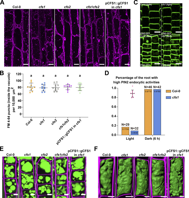
Figure 6.
Endocytic trafficking or vacuolar morphology is not affected in cfs1 mutants. (A) Confocal microscopy images of Arabidopsis root epidermal cells of Col-0, cfs1, cfs2, cfs1cfs2, and cfs1 complemented with pCFS1::gCFS1(pCFS1::gCFS1 in cfs1). 5-d-old Arabidopsis seedlings were first incubated in 4 μM FM 4-64-containing 1/2 MS media for 30 min and then transferred to 1 μM concanamycin A-containing 1/2 MS media for 2 h before imaging. Representative images of 10 replicates are shown. Scale bars, 10 μm. (B) Quantification of the FM 4-64 stained puncta inside the vacuole per normalized area (10,000 μm2) of the cells imaged in A. Bars indicate the mean ± SD of 10 replicates. One-way ANOVA tests were performed to analyze the differences of the number of FM 4-64 stained puncta between each group. Tukey’s multiple comparison tests were used for multiple comparisons. Family-wise significance and confidence level, 0.05 (95% confidence interval). (C) Representative microscopy images showing PIN2 endocytosis in the epidermal cells in the root tip meristem region of Col-0 and cfs1 under light or 6 h dark conditions. 5-d-old Arabidopsis seedlings expressing pPIN2::PIN2-GFP were grown on 1/2 MS media plates (+1% plant agar) under light or 6 h dark conditions before imaging. Scale bars, 10 μm. (D) Quantification of PIN2 endocytic activities in Col-0 and cfs1 shown in C. The Arabidopsis seedlings with at least five root epidermal cells that contained visible PIN2-GFP in the vacuole were considered as high PIN2 endocytic activities. The percentage of Col-0 and cfs1 with high PIN2 endocytic activities under light or 6-h dark conditions are shown in the graph. Numbers inside the bars represent the exact value (4 decimals) of each bar. N represents the total number of the Col-0 or cfs1 seedlings used for imaging and quantification in three independent experiments. (E) Confocal microscopy images showing the BCECF-AM-stained root epidermal cells in the meristem region of Col-0, cfs1, cfs2, cfs1cfs2, or pCFS1::gCFS1 in cfs1. 5-d-old Arabidopsis seedlings were incubated in 1/2 MS media containing 5 μM BCECF-AM for 30 min before imaging. Samples were mounted on slides with 0.002 mg/ml propidium iodide. Representative images of three replicates are shown. Green signals indicate the BCECF-AM-stained vacuole. Magenta signals indicate the propidium iodide-stained cell wall. Scale bars, 5 μm. (F) Three-dimensional images showing the vacuolar structure of the BCECF-AM-stained root epidermal cells in the transition region of Col-0, cfs1, cfs2, cfs1cfs2, or pCFS1::gCFS1 in cfs1. 5-d-old Arabidopsis seedlings were incubated in 1/2 MS media containing 5 μΜ BCECF-AM for 30 min before imaging. Samples were mounted on slides with 0.002 mg/ml propidium iodide. Representative images of three replicates are shown. Green signals indicate the BCECF-AM-stained vacuole. Magenta signals indicate the propidium iodide-stained cell wall. Scale bars, 10 μm.

