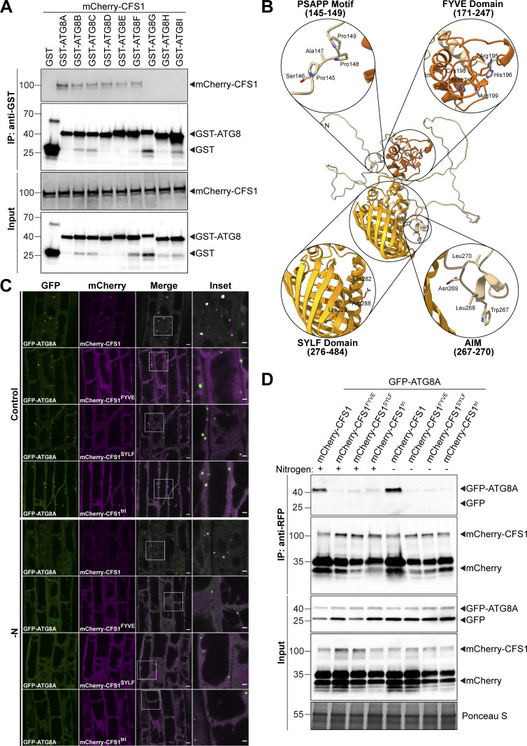
Figure S3.
Functional characterization of CFS1-ATG8 interaction. (A) GST pull-downs of E. coli lysates expressing either GST, GST-ATG8A, GST-ATG8B, GST-ATG8C, GST-ATG8D, GST-ATG8E, GST-ATG8F, GST-ATG8G, GST-ATG8H, or GST-ATG8I and A. thaliana whole-seedling lysates expressing mCherry-CFS1. Proteins were visualized by immunoblotting with anti-GST and anti-RFP antibodies. Representative images of two replicates are shown. Reference protein sizes are labeled as numbers at the left side of the blots (unit: kD). (B) Homology modeling and domain representation of CFS1. CFS1 structure is shown as ribbons, and relevant motifs and domains are highlighted as zoom-in, with the side chains of relevant residues represented as stick. For clarity, the FYVE and SYLF domains of CFS1 are colored in brick red and orange, respectively. (C) Confocal microscopy images of cfs1 mutants co-expressing pUBQ::GFP-ATG8A with either pUBQ::mCherry-CFS1, pUBQ::mCherry-CFS1FYVE, pUBQ::mCherry-CFS1SYLF or pUBQ::mCherry-CFS1tri. 5-d-old Arabidopsis seedlings were incubated in either control or nitrogen-deficient (−N) 1/2 MS media for 4 h before imaging. Representative images of 10 replicates are shown. Area highlighted in the white-boxed region in the merge panel was further enlarged and presented in the inset panel. Scale bars, 5 μm. Inset scale bars, 2 μm. (D) RFP-Trap pull-down of Arabidopsis seedlings co-expressing pUBQ::GFP-ATG8A with either pUBQ::mCherry-CFS1, pUBQ::mCherry-CFS1FYVE, pUBQ::mCherry-CFS1SYLF or pUBQ::mCherry-CFS1tri. 7-d-old seedlings were incubated in either control (+) or nitrogen-deficient (−) 1/2 MS media for 12 h. Protein extracts were immunoblotted with anti-GFP and anti-RFP antibodies. Representative images of two replicates are shown. Reference protein sizes are labeled as numbers at the left side of the blots (unit: kD). Source data are available for this figure: SourceData FS3.

