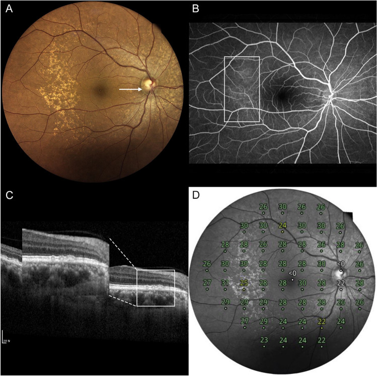
Figure 2.
A representative case of type 1 cuticular drusen. A 57-year-old woman presented with type 1 cuticular drusen. (A) Color fundus photography showed numerous cuticular drusen. The cuticular drusen area was scanned using spectral-domain optical coherence tomography (SD-OCT) (arrow). (B) Fundus fluorescein angiography showing hyperfluorescent drusen. This can confirm the diagnosis of cuticular drusen. (C) In the SD-OCT image scanned on the site of the arrow in Photo (A), the cuticular drusen area is magnified to categorize the types of cuticular drusen. It can be diagnosed as a type 1 cuticular drusen in which shallow elevation of the retinal pigment epithelium is present in a spectralis domain optical coherence tomography image. (D) Retinal sensitivity using microperimetry is topographically presented in a composited image.

