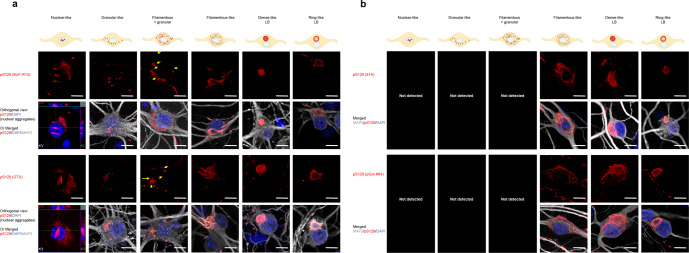
Fig. 5. All the pS129 antibodies do not detect the morphological diversity of the pS129-aSyn seeded-aggregates formed in human PFF-treated cortical neurons.
a, b After 21 days of treatment with 70 nM of human E83Q aSyn PFFs, primary cortical neurons were fixed, and ICC was performed. The seeded-aggregates were detected using the pS129 antibodies (MJF-R13, GTX, 81A, or pSyn#64). Neurons were counterstained with the MAP2 antibody and the nucleus with DAPI staining. Scale bar = 10 μM. All four pS129 antibodies were able to distinguish the seeded-aggregates with the filamentous-like morphology and the LB-like inclusions with the dense core and the ring-like morphology. However, only the MJF-R13 and GTX antibodies were able to detect the nuclear, granular or granular and filamentous-like aggregates (a). The yellow arrows indicate the filamentous-like aggregates imbricated between the granular-like aggregates. The orthogonal view from different planes (x/y, x/z, and y/z) of the “nuclear-like” pS129-aSyn aggregates stained with MJF-R13 or GTX antibodies as well as the Imaris 3D animations (see Videos in SI files) confirmed their intranuclear localization. The schematic representation of the different types of seeded-aggregates was created using BioRender.com.

