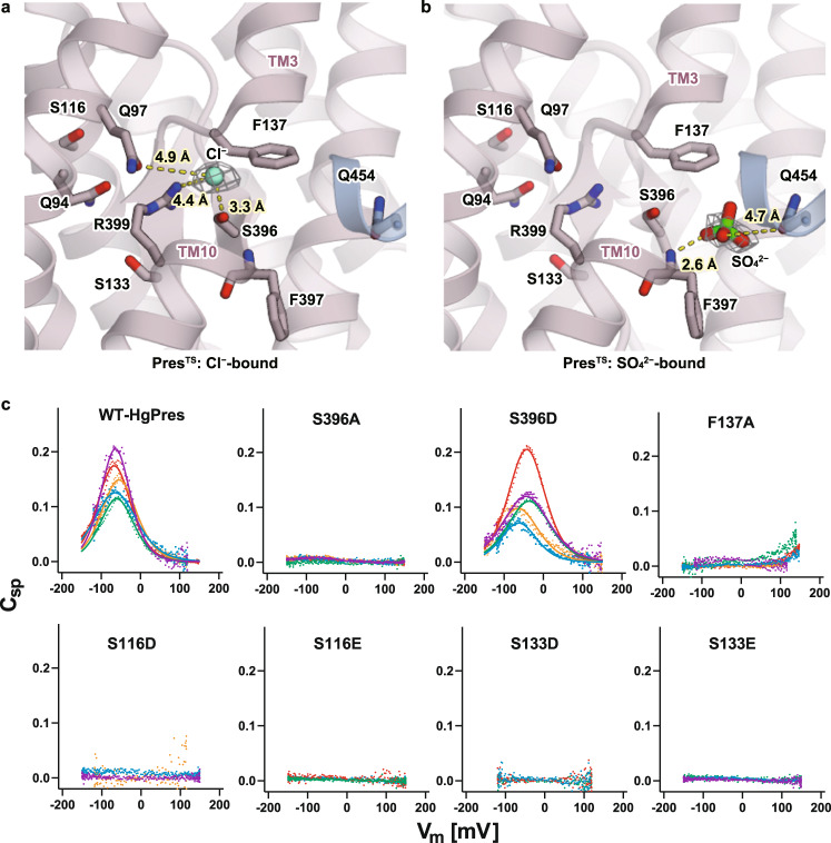
Fig. 2. The anion-binding sites of PresTS.
a, b Close-up views of the central anion-binding pocket found in the Cl−-bound (a) and SO42−-bound (b) PresTS structures. Cl− and SO42− ions are indicated as a green sphere and a stick model, respectively. Residues that contribute to anion binding are also shown with stick models. The cryo-EM densities of the bound anions are indicated by gray meshes. Predicted short-distance interactions are indicated by yellow dotted lines with the distance. c Cell membrane electric capacitance measurement in HEK293T cells expressing WT-, S396A-, S396D-, F137A-, S116D-, S116E-, S133D-, or S133E-HgPres. Three to five examples in different colors are shown for each panel. Solid lines for WT- and S396D-HgPres indicate two-state Boltzmann fittings. The α, Vpk, and charge density values (mean ± S.D.) were as follows: [0.032 ± 0.006 mV−1, −65 ± 14 mV, and 12 ± 5 fC/pF] for WT (n = 22); and [0.029 ± 0.005 mV−1, −51 ± 11 mV, and 14 ± 7 fC/pF] for S396D (n = 7). These NLC parameters were statistically indistinguishable between WT vs. S396D (determined by one-way ANOVA followed by Dunnett’s multi-comparison tests). Source data are provided as a Source Data file.

